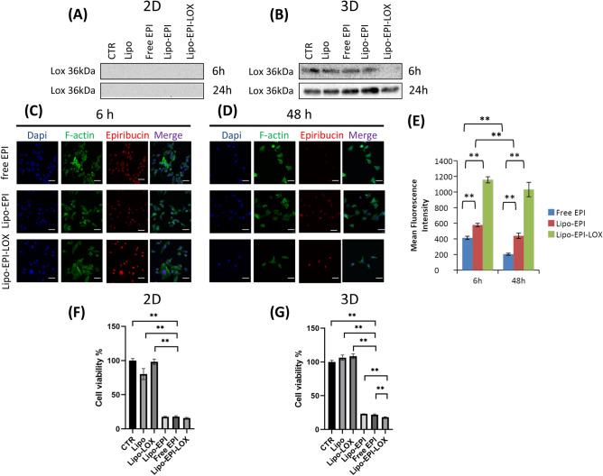
Figure 2.
(A) Protein analysis of secreted LOX in monolayer cultures not treated and treated with all formulations, full-length blots/gels are presented in Supplementary Fig. 2A. (B) Protein analysis of secreted LOX in 3D cultures not treated and treated with all formulations, full-length blots/gels are presented in Supplementary Fig. 2B, C. (C) Confocal analysis of 3D cultures exposed to formulations encapsulated with anthracycline after 6 h from the exposure. Nuclei were stained with dapi (blue), actin filaments were stained with phalloidin (green), epirubicin (red) and merge. (D) Confocal analysis of 3D cultures exposed to formulations encapsulated with anthracycline after 48 h from the exposure. Scale bar 50 µm (E) Mean fluorescence intensity of anthracycline detected after 6 and 48 h. (F) Cell viability of tumor cells after treatment with all studied formulations in standard monolayer cultures, negative control is CTR and positive control is LIPO-EPI-LOX. (G) Cell viability of tumor cells after treatment with all studied formulations in 3D cultures, negative control is CTR and positive control is LIPO-EPI-LOX. *p < 0.05, **p < 0.01.

