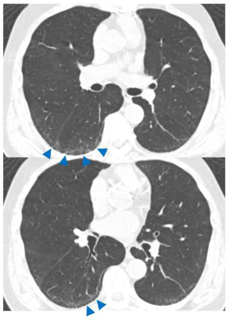
Figure 1.
TBI=1. CT images demonstrated subpleural ground-glass and reticular opacities indicating ILA. Note is made of dilatation of bronchioles (arrows) without obvious architectural distortion in the area of subpleural opacities of ILA. ILA, interstitial lung abnormalities; TBI, traction bronchiectasis index.

