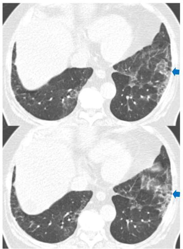
Figure 2.
TBI=2. CT images demonstrated ground-glass and reticular opacities with subpleural and basilar distribution indicating ILA. Note is made of mild bronchiectasis (arrows) associated with architectural distortion in the area of subpleural opacities of ILA. ILA, interstitial lung abnormalities; TBI, traction bronchiectasis index.

