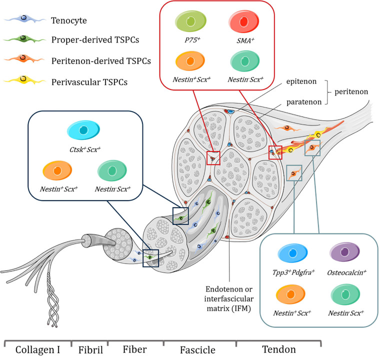
FIGURE 1.
Schematic representation of tendon hierarchical structure and various subpopulations of TSPCs with specific markers harvested from different niches, including tendon proper, peritenon and perivascular region. Tenocytes are aligned between fibers. It should be noted that some of these subpopulations might overlap with each other and perivascular TSPCs may be present in endotenon as well as the peritenon. What’s more, the exact location of proper-derived TSPCs is not well determined. Figures were produced using Servier Medical Art (https://smart.servier.com/).

