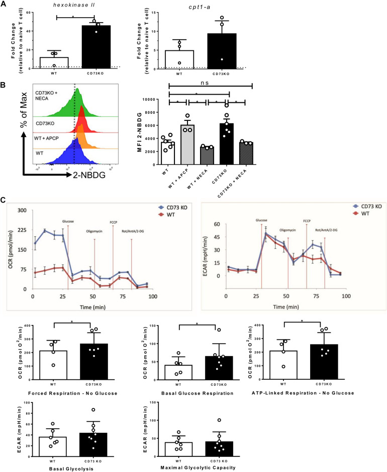
FIGURE 4.
CD73 restricts the mitochondrial capacity of Tc1 cells. Naive CD8+ T cells from WT mice were isolated and activated with soluble α-CD3/CD28 antibodies for 3 days in vitro in the presence of IL-2 (10 ng/ml) for the generation of Tc1 lymphocytes. (A) Expression of hexokinase II and cpt-1 mRNAs was analyzed by real-time PCR in CD73 and WT Tc1 cells (n = 3). (B) Glucose uptake of WT and CD73KO Tc1 cells treated with APCP (50 μM) or NECA (10 μM) was measured by FACS using the fluorescent glucose analog 2-NBDG (n = 3–6). (C) Left panel: Oxygen consumption rates (OCRs) and Right panel: extracellular acidification rates (ECARs) from CD73KO and WT Tc1 cells were measured under no glucose (forced mitochondrial respiration), basal glucose respiration and ATP-linked respiration for OCR and basal glycolysis and oligomycin stimulated glycolysis (maximal glycolytic capacity) for ECAR (n = 4;6 Tc1 WT and 8 CD73KO Tc1) Kruskal–Wallis Test, Paired T-test and Two-tailed Mann–Whitney test, *p < 0.05. Data is presented as the mean ± SEM.

