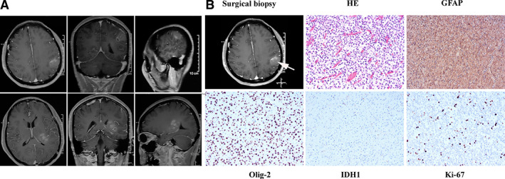
Figure 1.

Image of pathological diagnosis. (A): Magnetic resonance imaging revealed space‐occupying lesion in left thalamus and left parietal lobe. (B): Hematoxylin and eosin stain (200×) shows that tumor cells are dense and unevenly distributed, with obvious nuclear heterogeneity, and that small blood vessels proliferate in the tumor tissue. The immunohistochemical results showed that GFAP and Olig‐2 were expressed, IDH‐1 was negative, and the Ki67 proliferation index was ∼3%. The final diagnosis was anaplastic astrocytoma, IDH wild type, World Health Organization grade III. Abbreviations: GFAP, glial fibrillary acidic protein; HE, hematoxylin and eosin.
