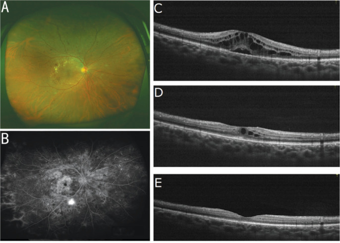
Figure 4. Fundus status of a patient with proliferative diabetic retinopathy before and after Conbercept treatment.

A: Fundus status before treatment; B: Fundus fluorescein angiography images before treatment; C: OCT examination images before treatment; D: OCT images after three successive Conbercept treatments; E: OCT images after 12 consecutive Conbercept treatments.
