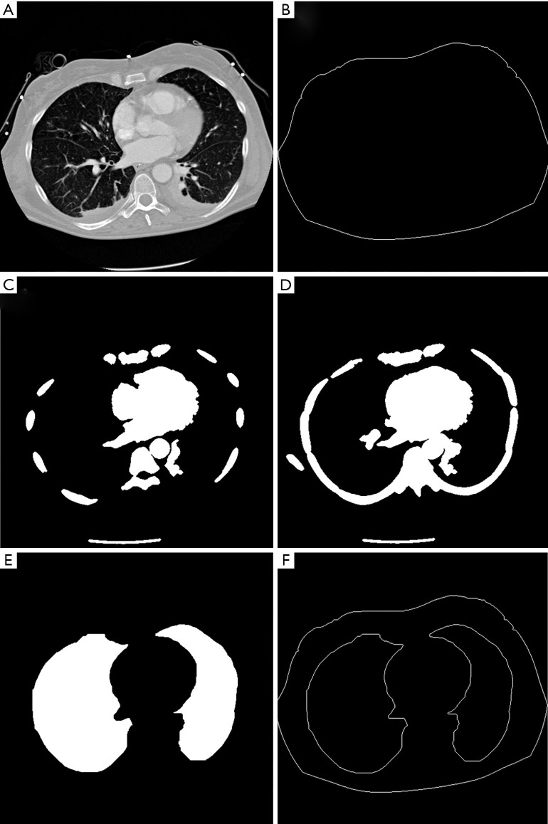
Figure 1.
Automatic segmentation of the thorax and lungs. (A) Serial CT images around the height of EIT electrode plane; (B) identified thorax contour; (C) ribs identified from one CT image; (D) ribs identified from a serial of CT images; (E) segmented lungs; (F) contours of thorax and lungs for further reconstruction.

