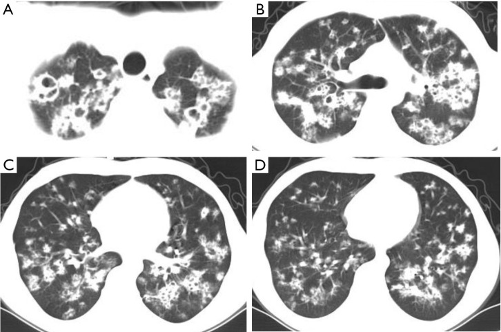
Figure 9.
A 26-year-old male patient with pulmonary tuberculosis. Lung CT shows diffusely distributed nodules and cystic changes in both lungs, and the lower lung air sacs are small and distributed in clusters. Thickening of the airway wall can be seen in the diseased area. A bronchoscopy lung biopsy revealed chronic inflammation and a small extent of necrosis of the bronchial mucosal tissue, and positive acid-fast staining. CT, computed tomography.

