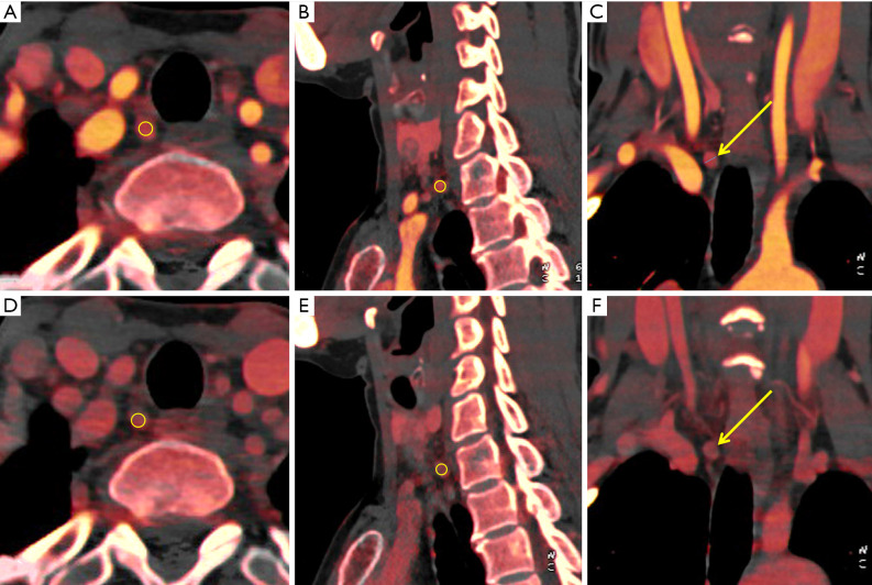
Figure 2.
A patient with PTC of the right lobe was confirmed by postoperative pathology, with metastatic lymph nodes in the right level VIb (3/3). (A,B,C) Iodine maps in the arterial phase. An ovoid region of interest (yellow; area, 9 mm2) was placed on the solid part in combined axial (A), sagittal (B), and coronal (C) images, including the entire lymph node as large as possible, and avoiding peripheral fat, cystic, necrosis, and calcification. (D,E,F) Iodine maps in the venous phase. The short diameter was 0.4 cm. In this case, the iodine concentration was 6.5 mg/mL in the arterial phase and 4.7 mg/mL in the venous phase. PTC, papillary thyroid carcinoma

