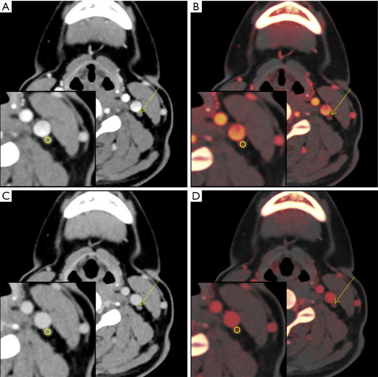
Figure 6.
Typical dual-energy CT images and iodine maps of non-metastatic lymph node with a diameter of 0.31 cm in a 37-year-old woman with PTC. (A) Contrast-enhanced monochromatic image in the arterial phase of a metastatic lymph node (area, 15 mm2, CT value, 40.2±7.2 HU). (B) Iodine map in the arterial phase of a metastatic lymph node (area, 15 mm2; iodine concentration, 0.8 mg/mL). (C) Contrast-enhanced monochromatic image in the venous phase of a metastatic lymph node (area, 15 mm2, CT value, 61.4±6.5 HU). (D) Iodine map in the venous phase of a metastatic lymph node (area, 15 mm2; iodine concentration, 1.3 mg/mL). CT, computed tomography; PTC, papillary thyroid carcinoma.

