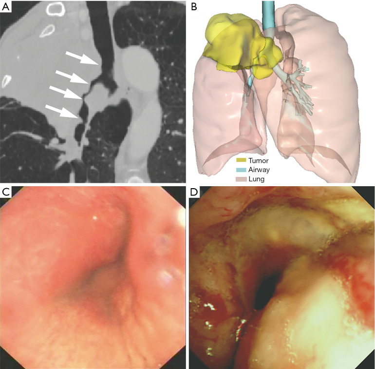
Figure 1.
A 59-year-old man with airway stenosis due to the invasion of LC. (A) Oblique coronal reconstructed CT images showed the narrow airway including the lower trachea, carina, RMB, and the BI (white arrow); (B) 3D reconstruction based on CT images showed the anatomical relationship between the airway and the tumor; (C,D) bronchoscopy showed an irregular neoplasm protruding into the carina and RMB. LC, lung cancer; CT, computed tomography; RMB, right main bronchus; BI, right main bronchus; 3D, three-dimensional.

