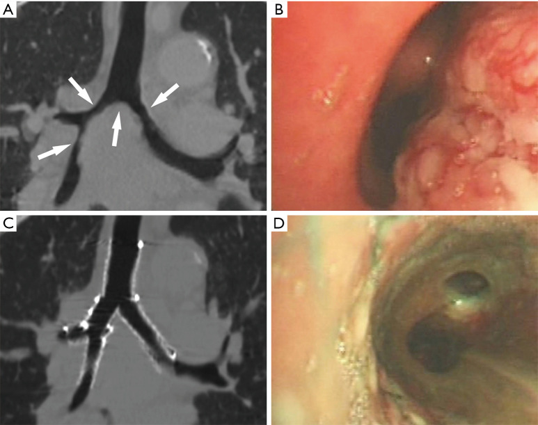
Figure 5.
CT and bronchoscopy images of the patient before and after double Y stenting. (A) Coronal reconstructed CT images showed the narrow airway including the carina, LMB, RMB, and the BI (white arrow); (B) bronchoscopy showed an irregular neoplasm protruding into the RMB; (C) coronal reconstructed CT images showed that the airway patency was restored and the 2 Y-shaped stents were in place; (D) bronchoscopy showed that the RMB was patent. CT, computed tomography; LMB, left main bronchus; RMB, right main bronchus; BI, right main bronchus.

