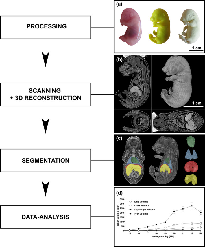
Figure 1.

Processing of fetal rat specimen, three‐dimensional reconstruction, and data analysis (a‐d). A native fetal rat (ED18; a left) was fixated in Bouin´s fluid (a center) and dried using CO2(“critical point drying”; a right). The dried specimen was scanned in the micro‐CT and the reconstructed data set was converted into a 3D model in sagittal, coronal, and transversal plane (b). Raw data sets were segmented from transversal sections into 3D models of single organs or structures using CTAn software leading to high‐resolution 3D models of single organs: heart (green), lung (blue), diaphragm (red), and liver (yellow) (c). Subsequently, total organ volumes of heart, lung, liver, and diaphragm from ED15 to N0 were calculated (d). All values presented as mean ± SD
