Figure 6.
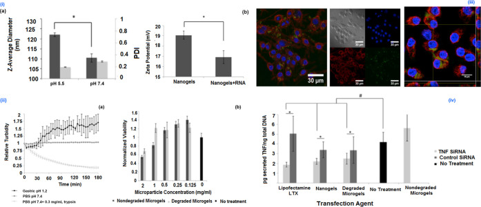
(i) (a) Hydrodynamic diameter of p(DEAEMA-co-tBMA) nanogels and (b) zeta potential measurements indicate a positive surface charge of ∼19 mV, which decreases slightly after loading with negatively charged RNA. (ii) (a) Relative turbidity measurements indicate that the microgel platform degrades in the presence of trypsin but remains intact in the presence of PBS or gastric fluid. (b) Relative viability of RAW 264.7 cells incubated with varying concentrations of degraded or nondegraded microgels for 24 h. (iii) (a) Bright-field/fluorescent panels and (b) Z-stack orthogonal images of RAW 264.7 macrophages incubated with degraded microgels containing P(DEAEMA-co-tBMA) nanogels. The released nanogels were efficiently uptaken by the RAW 264.7 cells (blue, DAPI; red, wheat germ agglutinin (membrane); green, NBD-Cl conjugated nanogels). (iv) TNF-α knockdown induced by siRNA carried by Lipofectamine LTX, p(DEAEMA-co-tBMA) nanogels, or degraded microgels containing p(DEAEMA-co-tBMA) nanogels. Reprinted with permission from ref (12). Copyright 2016 American Chemical Society.

