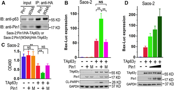
Fig. 1.

Pin1 enhances TAp63γ‐induced transcription and apoptosis. (A) Saos‐2 cells transfected with HA‐TAp63γ, plus Pin1 or its W34A mutant, were lysed and subjected to IP with anti‐HA. The cell lysates (inputs) or IP products were subjected to immunoblot (IB) analysis with indicated primary antibodies. (B) Saos‐2 cells were transfected with a mixture of Bax‐Luc and TK‐Renilla plus indicated plasmids. M, W34A mutant Pin1. Firefly and Renilla luciferase activities were measured, while IB analyses were performed to detect indicated proteins. The Bax‐Luc activity was normalized to Renilla activity and presented as Bax‐Luc expression level with SD (n = 3). Bax‐Luc expression in cells transfected with Bax‐Luc/TK‐Renilla mixture alone was set as 1. Two‐tailed t‐test was used for comparison between two groups; **P < 0.01; NS, nonsignificant. (C) Saos‐2 cells transfected with indicated plasmids were subjected to cell survival measurement with 3‐(4,5‐dimethylthiazol‐2‐yl)‐2,5‐diphenyl‐tetrazolium bromide (MTT). Cell viabilities were presented as optical density values at the wavelength of 490 nm (OD490) with SD (n = 3). Two‐tailed t‐test was used for comparison between two groups; **P < 0.01; NS, nonsignificant. (D) Saos‐2 cells were transfected with a mixture of Bax‐Luc and TK‐Renilla plus HA‐TAp63γ and increasing amounts of Pin1 plasmid as indicated. Bax‐Luc expression levels were measured and presented as mentioned above, while IB analyses were performed to detect indicated proteins. The error bars indicate SD (n = 3).
