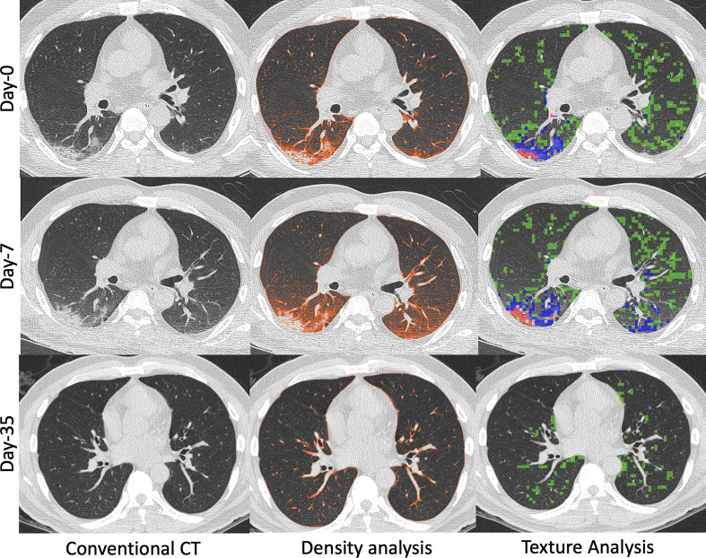
Figure 2.
55-year-old male with COVID-19 pneumonia that received imaging at presentation (Day 0), Day 7 of hospitalization, and Day 35 of hospitalization. Images generated at initial presentation (Top row, Day 0) along with computed density and texture analysis demonstrating predominantly posterior bilateral lower lobe disease (HAAs quantified as 13% on density analysis and abnormal lung quantified as 24% on texture analysis). The texture analysis characterized the disease as GGO (green, 21%), mixed GGO and interstitial (blue, 3.9%), and consolidation (red, 0.1%). Follow-up CT (Middle row, Day 7) obtained due to worsening shortness of breath demonstrated minimal disease progression on conventional CT images and density analysis and texture analysis provided objective assessments of disease worsening (HAA on density analysis quantified as 18% and abnormal lung quantified as 29% on texture analysis). Progression of imaging disease type was also quantified (GGO, green: 22.5%; mixed GGO and interstitial, blue: 6.3%; and consolidation, red: 0.2%). Another follow-up CT (Bottom row, Day 35) obtained after clinical improvement and prior to discharge from the hospital demonstrated complete resolution of disease on conventional images. The density and texture analysis identified some residual abnormalities (HAA 5% on density analysis and abnormal lung 6% on texture analysis). GGO, ground glassopacity; HAA, high attenuation area

