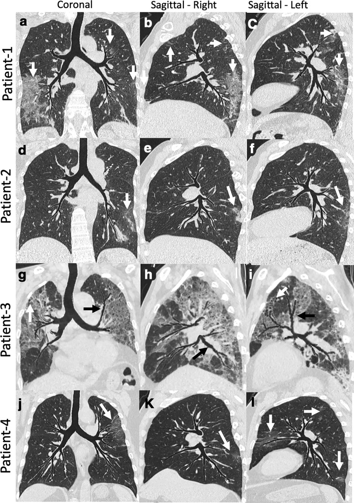
Figure 3.
tMPR view images of four patients with COVID-19 pneumonia. The tMPR view warps the airway tree such that most major subsegmental airway segments reside within a single flattened projection, and associated parenchyma and vasculature are similarly warped. Patient 1: Coronal (a) and Sagittal (b, c) images showing multifocal, predominantly peripheral bilateral ground glass opacification (white arrows). These images provide comprehensive visualization of airway and overall extent of disease. Patient 2: Coronal (d) and sagittal (e, f) images showing bilateral peripheral mixed ground glass opacity and reticular thickening (white arrows). The coronal image demonstrates left-sided disease, but the right sagittal image demonstrates posterior lower lobe disease as well. Patient 3: Coronal (g) and sagittal (h, i) images demonstrating extensive bilateral upper and lower lobe disease. The patient had a protracted course and the imaging was obtained later in the time course. Images show bilateral central bronchiectasis (black arrows) and distal bronchiolectasis (white arrow). Patient 4: Coronal (j) and sagittal (k, l) images demonstrate multifocal (white arrows) left-side-predominant disease. Review of these three images provide a good appreciation of localization and the relationship with the airway paths. tMPR, topographic multiplanar reformatted.

