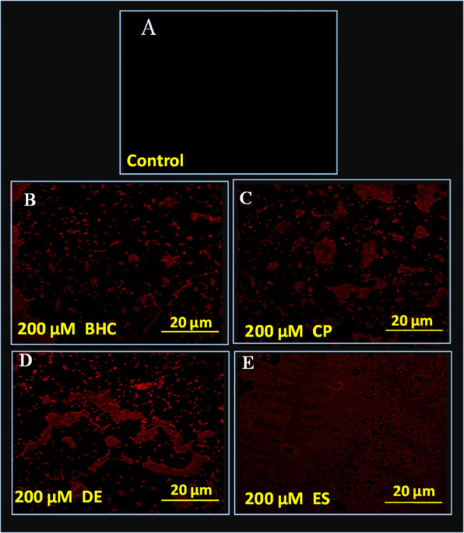
Figure 5.

PI-stained confocal laser scanning microscopy images of E. cloacae strain EAM 35: panel (A) shows untreated control cells with no red rod-shaped cells, whereas panels (B, C, D, and E) represent the red rods treated with 200 μM each benzene hexachloride, chlorpyrifos, dieldrin, and endosulfan. In these images, red rods depict membrane-compromised cells.
