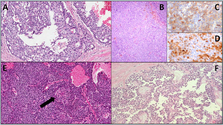
Figure 4.

(A) Representative microscopic image of an acinar cell carcinoma, with aspects of intraductal growth (original magnification 20X); (B,C,D) Representative images of a mixed neuroendocrine-acinar neoplasm: B: hematoxylin-eosin staining (original magnification 10X), C: immunohistochemistry for synaptophysin (original magnification 20X), D: immunohistochemistry for trypsin (original magnification 20X); (E) representative histological image of a pancreatoblastoma, including a squamous nest, indicated with a black arrow (original magnification 20X); (F) representative microscopic image of a solid-pseudopapillary neoplasm (original magnification 10X).
