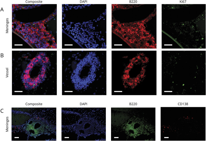
Figure 2. Plasma Cells and Proliferating B Cells Can Be Observed in DTH-TLS Lesions.
Focal MS-like DTH-TLS lesions were established in mice. Brains were stained for B cells (B220 in red), proliferation (Ki67 in green), and DAPI (blue) using immunofluorescence. The DTH-TLS model features proliferating B cells in both the meninges (A) and in vessel cuffs (B). CD138+ plasma cells were also present in the lesions (in red), outside of the area occupied by B cells (in green) (C). White scale bars represent 100 μm. TLS = tertiary lymphoid structures.

