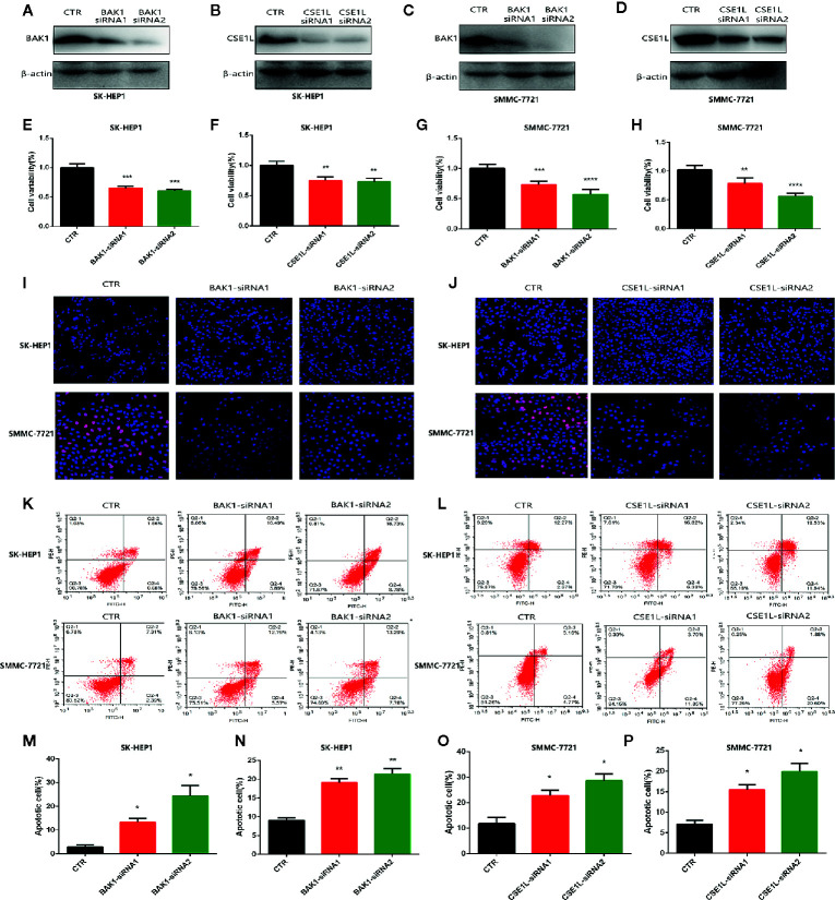
Figure 2.
Measure the oncogenic effect of BAK1 and CSE1L in the HCC. (A–D) Western blot determined that expression of BAK1 and CSE1L was inhibited with BAK1 and CSE1L siRNA administration. (E–H) CCK8 assay showed BAK1 and CSE1L inhibition significantly suppressed HCC cells proliferation. (I, J) Edu assay indicated knock down BAK1 and CSE1L inhibited HCC cells proliferation, respectively. (K, L) Flow cytometry confirmed decreased BAK1 and CSE1L expression promoted HCC cells apoptosis in the HCC. (M–P) Quantitative statistical results of the effects of BAK1 and CSE1L expression on cell apoptosis. Data are shown as the mean ± SD of at least three independent experiments. *P < 0.05, **P < 0.01, ***P < 0.001, ****P < 0.0001.

