FIGURE 1.

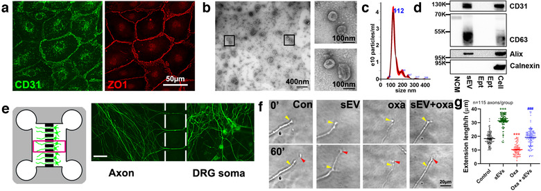
CEC‐sEVs promote axonal growth of DRG neurons with the presence of oxaliplatin. [Representative confocal microscopic images (a) show CECs are CD31 (green, CD31) and ZO1 positive (red, ZO‐1). Characterization of CEC‐sEVs by TEM (b), NTA (c) and Western blots (d), respectively. Schematic figure of the standard microfluidic device (SND150) along with an immunofluorescent image captured in the box area (e) shows DRG neurons grown in the cell body compartment (DRG soma) and their axons in 150 μm long microgrooves and in the axonal compartment (Axon). Representative time‐lapse microscopic images (f) of growth cone extension within 60 min and corresponding quantitative data of growth cone extension during a 24 h period (g), respectively, under control (con), CEC‐sEVs (sEV), oxaliplatin (oxa) and CEC‐sEVs in combination with oxaliplatin (sEV+oxa) conditions. Yellow and red arrows in panel G indicate the start (0′) and end positions (60′), respectively. One‐way ANOVA with Tukey's multiple comparisons test was used. *** P < 0.001 vs. control. N indicates the number of axonal growth cones. In panel D, NCM = the particles isolated from non‐conditioned medium, sEVs = CEC‐sEVs, Ept = the intentionally empty lanes, Cell = CEC lysate, K = the molecular weight Kda. Error bars indicate the standard error of the mean (SEM)]
