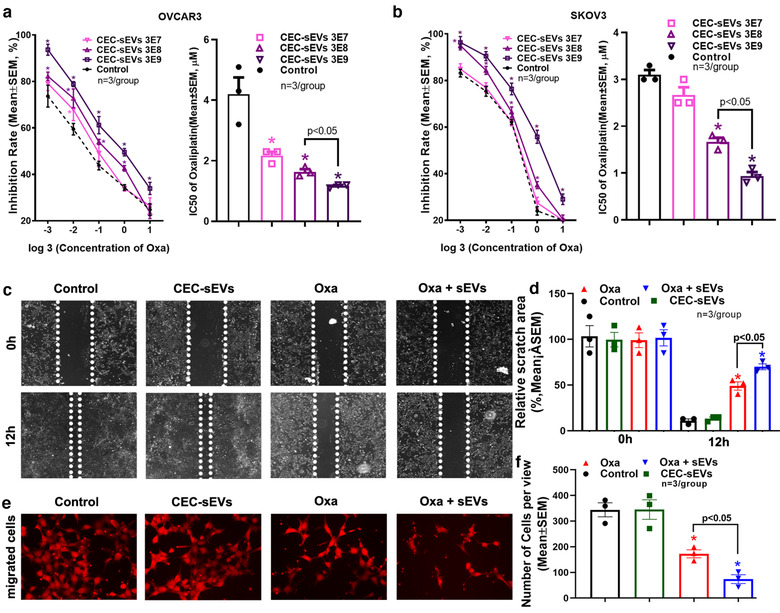
FIGURE 2.

CEC‐sEVs enhanced anti‐cancer effects of oxaliplatin in OC cells. [Quantitative data of MTT cell viability assays on OVCAR3 (a) and SKOV3 (b) cells show the inhibition rates and corresponding IC50, respectively, of oxaliplatin in combination with different concentrations of CEC‐sEVs. Representative images (c) and quantitative data (d) show the results of a 12 h‐period wound healing assay of OVCAR3 cells treated with PBS (control), CEC‐sEVs, oxaliplatin (oxa) and CEC‐sEVs in combination with oxaliplatin (oxa + sEVs). Representative images (e) and quantitative data (f) show the results of the Transwell migration assay of OVCAR3 cells treated with different conditions for 24 h. N indicates the replications. One‐way ANOVA with Tukey's multiple comparisons test was used. * P < 0.05 vs. control; Error bars indicate the standard error of the mean (SEM)]
