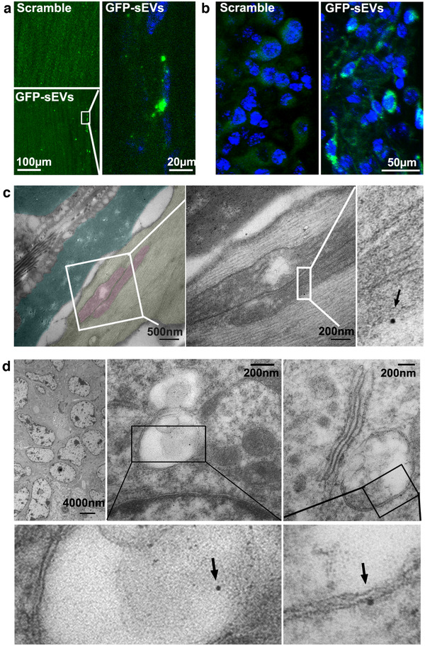
FIGURE 7.

The internalization of CEC‐sEVs in sciatic nerves and OC tumor cells. [Confocal microscopic images show the presence of GFP signals in sciatic nerve fibres (a) and OC tumor cells (b) with the administration of GFP‐sEVs in nude mice bearing OC tumors. Representative TEM images with enlarged areas (c) show the GFP positive gold particle (black arrows) is associated with the mitochondria (pink) of damaged axons (yellow) with separated myelin sheath (dark green). Representative TEM images with enlarged areas (c) show the GFP positive gold particles (black arrows) are associated with MVBs in OC tumor cells]
