Figure 1.
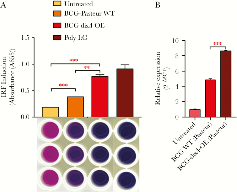
BCG-disA-OE overexpressing bis-(3’-5’)-cyclic dimeric adenosine monophosphate (c-di-AMP) gives more potent interferon (IFN) regulatory factor (IRF) 3 and type I IFN stimulation than bacillus Calmette-Guérin (BCG)-wild-type (WT): (A) effect of disA overexpression on activation of IRF pathway measured by IRF- secreted embryonic alkaline phosphatase (SEAP) QUANTI Blue reporter assay. RAW-Blue IFN-stimulated gene (ISG) cells were challenged with WT and BCG-disA-OE strains at a multiplicity of infection (MOI) of 1:20 for 5 hours to establish the infection. Uninfected bacteria were washed out using ice-cold Dulbecco’s phosphate-buffered saline (DPBS), and the RAW-Blue ISG cells were subsequently incubated for another 18–24 hours. The culture supernatants of infected RAW-Blue ISG cells were assayed for IRF activation. The image below the IRF-activation graph represents QUANTI Blue assay plate and sample wells; treatment parameters for column of wells correspond to those defined for the bars above aligned with the wells. The graphical points represent the mean of 3 independent experiments ± standard error of mean (SEM). Student’s t test (***, P < .001; **, P < .01). (B) Differential expression of IFN-β: mouse bone marrow-derived macrophages were challenged with WT and BCG-disA-OE strains at an MOI of 1:20 for 5 hours to establish the infection. Uninfected bacteria were washed using ice-cold DPBS, and cells were subsequently incubated for another 6 hours. Expression levels of messenger ribonucleic acid (mRNA) were measured using a SYBR Green-based quantitative real-time polymerase chain reaction (PCR). Basal level of transcript (mRNA) in untreated macrophages was used for data normalization and hence to access relative expression. β-actin was used as an internal control. Data analysis was performed using the 2−∆∆CT method. The graphical points represent mean of 3 independent experiments ± SEM. Mean comparison between BCG WT and BCG-disA OE were performed by Student’s t test (***, P < .001).

