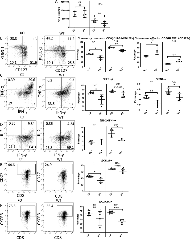
Figure 7. Zbtb20 deletion affects effector CD8 T cell differentiation during acute LM infection.
Naïve CD8 T cells were harvested from CD45.1 OT-I mice (WT) or GZB-cre Zbtb20-fl/fl CD45.1 OT-I mice (KO). Naïve OT-I cells were retro-orbitally injected into B6 recipients, which were then retro-orbitally infected with LM-actA-Ova 1 day later. Splenocytes were harvested on day 7 or 14 post-infection and analyzed by flow cytometry. (A-F) All plots were gated on transferred OT-I cells. (A) Total cell counts for transferred OT-I cells from the spleen of each recipient. (B) Representative dot plot showing KLRG-1 and CD127 staining to measure memory precursor cells (KLRG-1-CD127+) and terminal effector cells (KLRG-1+CD127-), and quantification. (C) Representative dot plot showing TNF-α and IFN-γ staining and quantification. (D) Representative dot plot showing IL-2 and IFN-γ staining and quantification. (E) Representative dot plot showing CD27 and CD8 staining and quantification. (F) Representative dot plot showing CXCR3 and CD8 staining and quantification. Each point represents data from an individual mouse. Each group used at least four mice and each experiment was repeated three times. Numbers in dot plot quadrants show percentage of events in the relevant quadrant. Statistics were performed with Student’s unpaired t-tests. *P<0.05, **P<0.01, ***P<0.001.

