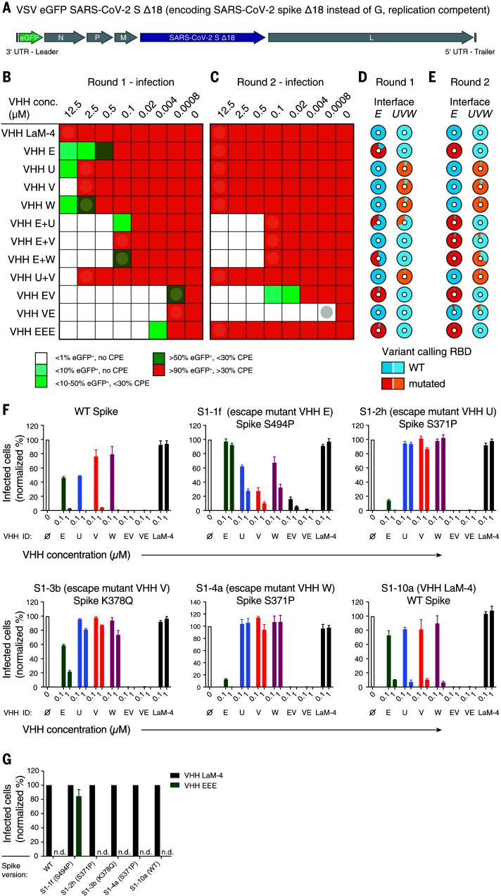
Fig. 5. Simultaneous targeting of two independent epitopes with neutralizing VHHs prevents viral escape.
(A) Genome structure of VSV SARS-CoV-2 S Δ18 eGFP. UTR, untranslated region. (B to E) Evolution experiment. (B) Replication-competent VSV SARS-CoV-2 S Δ18 eGFP at a multiplicity of infection (MOI) of 0.5 was incubated with different concentrations of the indicated VHHs and allowed to replicate on Vero E6 cells in 12 wells for 4 days. The fraction of infected (eGFP-positive) cells and the cytopathic effect (CPE) were estimated microscopically and are plotted according to the indicated color code. (C) Cleared supernatants from the wells indicated with a circle in (B) were diluted with four volumes of fresh infection medium (1:5 dilution) and used for a second round of replication on Vero E6 cells in the presence of the indicated VHH concentrations. Cleared supernatants were harvested as in (B). (D and E) Cell lysates from the wells indicated by circles in (B) and (C) [corresponding to (D) and (E), respectively] were subjected to targeted resequencing of the RBD to identify variants that had emerged at the interfaces to VHH E (interface E) or to VHH U, V, or W (interface UVW) and to quantify their allelic distribution (see tables S5 and S6 for details on detected variants). (F and G) Wild-type VSV SARS-CoV-2 spike eGFP or plaque-purified escape mutants of VHH E (S1-1f, Spike S494P), VHH U (S1-2h, Spike S371P), VHH V (S1-3b, Spike K378Q), VHH W (S1-4a, Spike S371P), and VHH LaM-4 (S1-10a, WT spike) at an MOI of 0.5 were incubated with the indicated VHH concentrations (F) or 1 μM of the indicated VHH (G) and used for Vero E6 infection experiments as in Fig. 1B. Infection was quantified by flow cytometry; normalized data from three independent experiments ± SEM are plotted. n.d., not detected.

