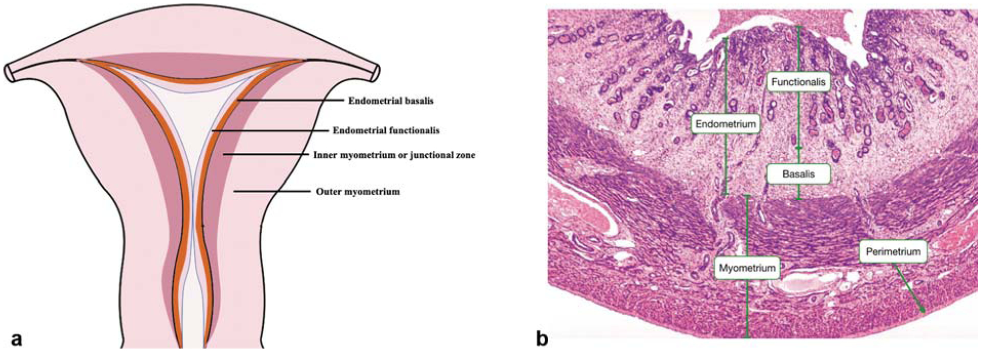
Fig. 1. The structure of normal uterus.

(a) The coronal section of normal uterus. (b) Hematoxylin–eosin staining of the full-thickness of uterus. (With permission from Yale Histology. Available at: http://histology.med.yale.edu/female_reproductive_system/female_reproductive_system_reading.php.)
