
Figure 1.
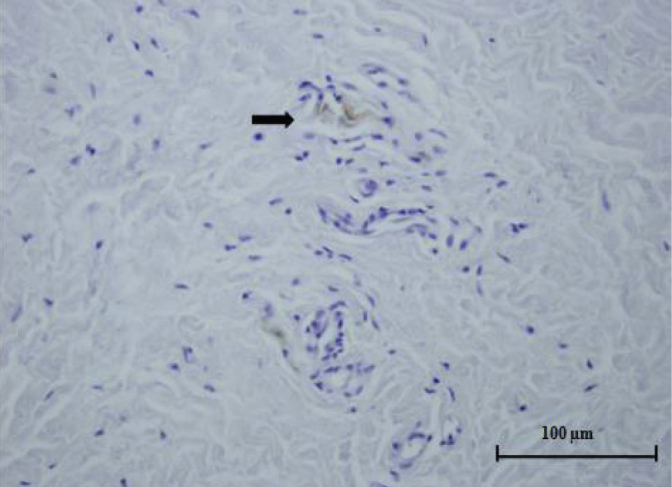
Histological specimen from the pulvinar. Free nerve ending (arrow) can be seen in the fibrovascular connective tissue (light microscope x400, S-100 protein)
Official websites use .gov
A
.gov website belongs to an official
government organization in the United States.
Secure .gov websites use HTTPS
A lock (
) or https:// means you've safely
connected to the .gov website. Share sensitive
information only on official, secure websites.
Histological specimen from the pulvinar. Free nerve ending (arrow) can be seen in the fibrovascular connective tissue (light microscope x400, S-100 protein)