
Fig. 6—
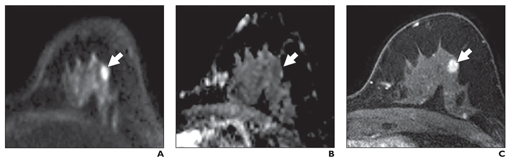
58-year-old woman with dense breasts and mammographically occult invasive ductal carcinoma. Patient underwent high-risk screening MRI (3 T) because of strong family history of breast cancer. On DWI, this lesion received unblinded visual conspicuity score of 5 and was correctly identified in blinded reader study by two of three readers.
A, Axial DW image (b value = 800 s/mm2) shows area of high signal intensity (arrow).
B, Apparent diffusion coefficient (ADC) map shows low diffusivity of lesion (arrow). ADC value is 1.45 × 10⁻³ mm2/s.
C, T1-weighted dynamic contrast-enhanced MR image (provided here for reference, not provided to readers) shows enhancing 8-mm mass (arrow).