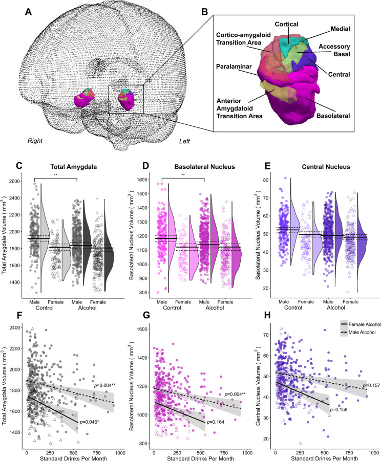
Fig. 1. Overview of group by sex effects on the volume of a-priori amygdala region and amygdala nuclei.
A Overview of glass brain section showing 3D rendering and B close up of 3D renders of all amygdala nuclei from an example participant. Plots of the a priori amygdala regions, comprising C total amygdala in greyscale, D basolateral nuclei in pink, and E central nuclei in purple containing individual data points and the probability density of the data stratified by group and sex (the group average of the estimated marginal means predicted by the models are indicated by the solid black line, with dotted lines indicating the standard error of the marginal means). The bottom panel shows regression plots for volume of the F total amygdala, G basolateral nuclei, and H central nuclei by alcohol use (standard alcohol drinks per month) in male and female alcohol-dependent participants, adjusted for intracranial volume, age, and education. Only the association in the male group for the total and basolateral amygdala remained significant after FDR correction. The left and right hemispheres for the nuclei have been collapsed. **p(FDR) < 05.

