Figure 5.
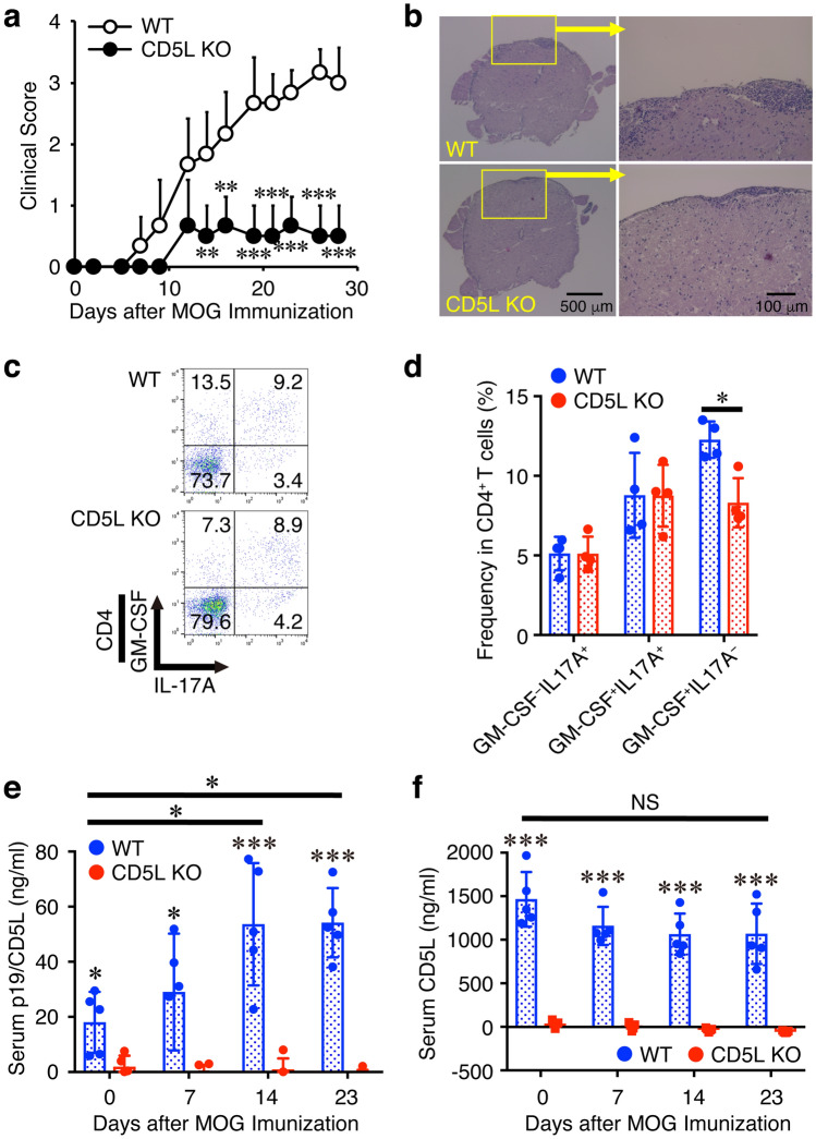
CD5L-deficient mice show alleviated EAE with reduced frequency of GM-CSF+CD4+ T cells in the CNS. (a–d) WT mice or CD5L-deficient mice were immunized with MOG35-55 peptide and their clinical scores were monitored with time (a). On day 15, spinal cords and brains were harvested, and the CNS was histopathologically analyzed with H&E staining. Representative images are shown (b). Mononuclear cells were also isolated from the CNS, and intracellular cytokine staining was performed after restimulation with PMA and ionomycin. Representative dot plots of GM-CSF, and IL-17A in CD4+ T cells are shown (c). Average frequencies of respective CD4+ T cells were calculated and compared (d). Blood was also taken over time and serum levels of p19/CD5L and CD5L were determined by ELISA (e, f). Data are shown as mean ± SD (n = 4–6) and are representative of three independent experiments. P values were determined using unpaired, two-tailed Student’s t-test (a, d) or one-way ANOVA (e, f). *P < 0.05, **P < 0.01, ***P < 0.001. NS, not significant.

