
Figure 7.
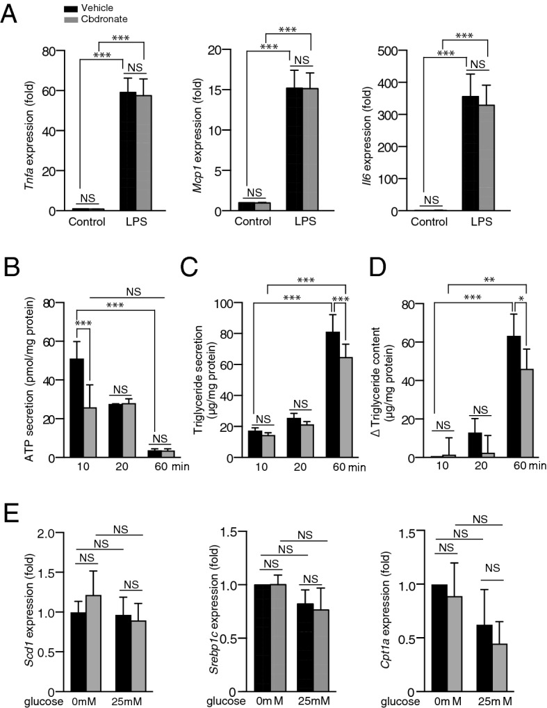
Clodronate inhibits ATP release and triglyceride synthesis but not inflammatory gene expression in mouse primary hepatocytes. Primary hepatocytes were isolated from 10-week-old C57BL/6 male mice. (A) Primary mouse hepatocytes were pre-incubated in the presence or absence of 10 µM clodronate for 30 min, followed by stimulation with 1 µg/mL lipopolysaccharide (LPS) or the control (saline) added to the medium. The mRNA levels of the inflammatory cytokine genes, Tnfa, Mcp1, and Il6 in the mouse primary hepatocytes were determined 4 h after stimulation by qRT-PCR. (B–D) Primary hepatocytes were pre-incubated for 20 min with glucose-free Krebs–Ringer bicarbonate buffer with or without 10 µM clodronate, followed by the administration of Krebs–Ringer buffer containing 25 mM glucose with or without 10 µM clodronate for the indicated time. The amounts of ATP (B) and triglycerides (C) in the culture supernatants, as well as the triglyceride contents in the primary hepatocytes (D), were evaluated at the indicated time points. The change in triglyceride content (D) was calculated based on the increase from the baseline value (hepatocytes treated with Krebs–Ringer buffer containing 25 mM glucose without clodronate at the time point of 10 min). (E) The mRNA expression levels of Scd1, Srebp1c, and Cpt1a were analyzed by qRT-PCR 60 min after either 0 mM or 25 mM glucose stimulation. Gene expression is shown as the fold change relative to hepatocytes treated with glucose-free Krebs–Ringer buffer. All data are shown as the mean ± S.E.M. *P < 0.05, **P < 0.01, ***P < 0.001. NS not significant.