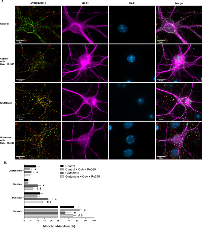
Figure 4.
Morphological analysis of mitochondria in glutamate and cyclosporin A (CsA) + Ru360 experiments. (A) Representative images of primary cortical neurons. Rows (top to bottom): vehicle-treated control group, control cells treated with CsA + Ru360 only, vehicle-treated group with 30 min 100 µm glutamate exposure, glutamate challenged group pre-treated and co-incubated with CsA + Ru360 Panels (from left to right): ATP synthase (green) and TOM20 (red) merged, MAP2 (magenta), DAPI (blue), all channels merged. Scale bars = 10 µm. (B) Percentage of mitochondrial area per morphology in glutamate and CsA + Ru360 experiments. Mean ± SD (n = 6–8 per group). * indicates p < .05 versus Control. # indicates p < .05 versus Control + CsA + Ru360. $ indicates p < .05 versus Glutamate.

