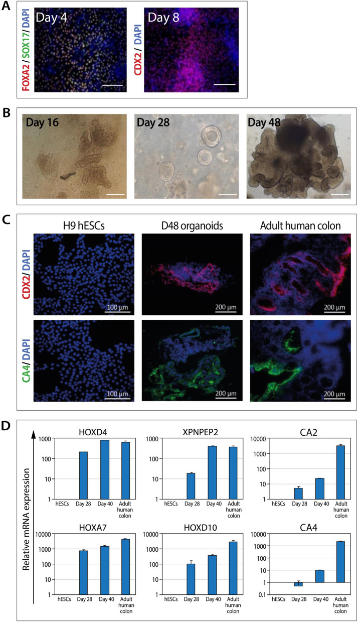
Figure 1.
Characterization of hESC-derived colon organoids. (A) Immunostaining of H9 hESC-derived definitive endoderm (day 4) and hindgut endoderm (day 8). Scale bars: 200 µM. (B) Representative bright-field images of developing hESC-derived colon organoids. Scale bars: 200 µM. (C) Immunostaining of hESC-derived colon organoids. Adult human colon tissue samples and H9 hESCs served as controls. Scale bars: 200 µM. (D) RT-PCR analysis of colonic cell markers in hESC-derived D28 colonic spheroids and D40 colon organoids. Adult human colon tissue samples and H9 hESCs served as positive and negative controls, respectively. Data are presented as relative mean expression (± standard deviation) of triplicate samples from one experiment.

