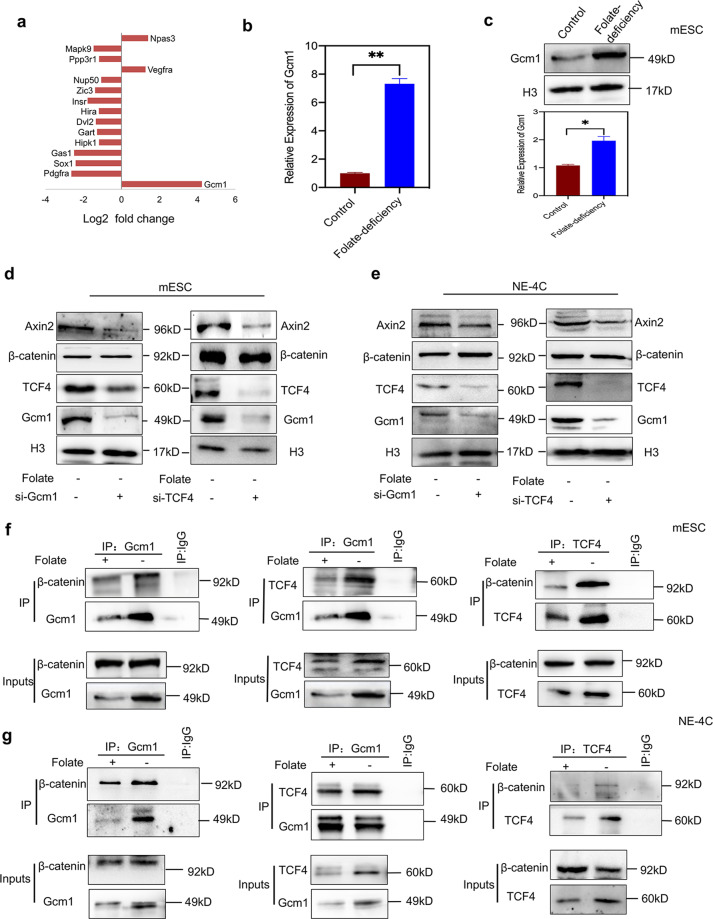
Fig. 2. Folate deficiency upregulates Gcm1 to promote formation of the Gcm1/β-catenin/TCF4 complex.
a Differentially expressed NTD-related genes in the microarray expression profiles of C57BL/6 mESCs cultured in folate-deficient conditions for six generations. The microarray data were obtained from three replicates for both control and folate-deficient cells. The X-axis represents the Log2 fold change. bThe mRNA expression of Gcm1 in C57BL/6 mESCs after six generations of folate deficiency. **P = 0.004. c Increased expression of Gcm1 in mESCs with folate deficiency. Top panel: western blots of Gcm1 in control and folate-deficient cells; bottom panel: bar graph showing the quantification of the western blot signal intensities. d Efficiency of siRNA-mediated Gcm1 and siRNA-mediated TCF4 depletion and the expression of TCF4, Gcm1 and Axin2 at the protein level in C57BL/6 mESCs. mESCs with folate deficiency were transfected into either scramble siRNA control or Gcm1 siRNA and TCF siRNA, and post 72 h of transfection, the expression was determined by western blotting. e Efficiency of siRNA-mediated Gcm1 and TCF4 depletion and the expression of TCF4 and Axin2 at the protein level in NE-4C. NE-4C with folate deficiency were transfected into either scramble siRNA control or Gcm1 siRNA, TCF4-siRNA, and the expression was determined 72 h post transfection by western blotting. f A coimmunoprecipitation (Co-IP) assay was used to identify the interaction between Gcm1, β-catenin, and TCF4 in C57BL/6 mESC. Immunoprecipitation (IP) was performed using an anti-Gcm1 antibody and TCF4 antibody, and immunoblotting (IB) was performed using an anti-β-catenin antibody. g A Co-IP assay was used to identify the interactions between Gcm1 and TCF4, β-catenin in NE-4C. An anti-Gcm1 antibody and anti-TCF4 antibody was used for IP, and an anti-TCF4 antibody and anti-β-catenin was used for IB. Data b–f represent the mean ± SEM (n = 3). The p value was calculated by Student’s t-test, ns was for no significance, *P < 0.05, **P < 0.01, ***P < 0.001.

