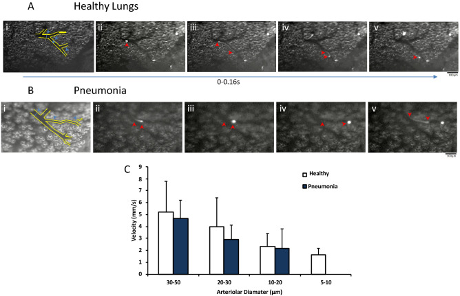
Figure 3.
Real-time Intra-vital recording of MSC accumulation in the murine lung microvasculature allowed for measurements of velocity in healthy (A) and E. coli infected lung (B). Velocities of individual MSCs (movement indicated by red arrows, A, B, Panels ii–v) were measured in different pulmonary vessel diameters (Blue arrows, A,B, Panel i) Quantitative analysis showed that there was no significant differences in MSC velocities in different vessel diameters between healthy and pneumonia mouse models (C). MSC: Mesenchymal Stem Cell; Columns represent mean + SD (n = 4–13 from 6 separate animals).

