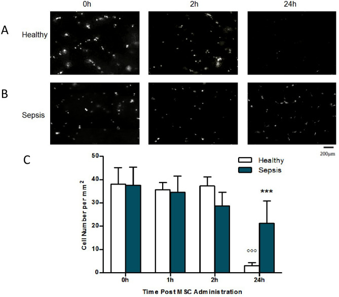
Figure 5.
IVM was performed at 0 h, 2 h and 24 h post fluorescent MSC administration and composite images rendered from lung surface recordings (A,B). Quantitative analysis of cell numbers in composite images demonstrated no differences in cell retention between healthy and septic animals from 0 to 6 h (C). However, a significantly higher number of cells remained in animals with E. coli pneumonia 24 h post cell administration (A,B,C). IVM: Intravital Microscopy; MSC: Mesenchymal Stem Cell; Columns represent mean + SD (n = 2–5), * = P < 0.05 versus 24 h Healthy, o = P < 0.05 versus 0 h Healthy.

