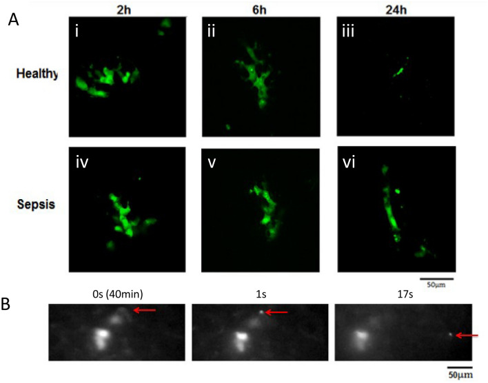
Figure 6.
In vivo confocal microscopy allowed detailed analysis of cell structure at each time- point in healthy and pneumonia lungs with cell size differences discernibly evident at 24 h between MSCs in healthy and pneumonia lungs. The formation of micro-particles from the cells was indicated in post mortem confocal microscopy (A) and performing IVM using a 40X objective demonstrated the time-lapse release of fluorescent micro-particles (B, Red Arrow) approximately 40 min post cell administration IV (B i-iii) IVM: Intravital Microscopy; MSC: Mesenchymal Stem Cell.

