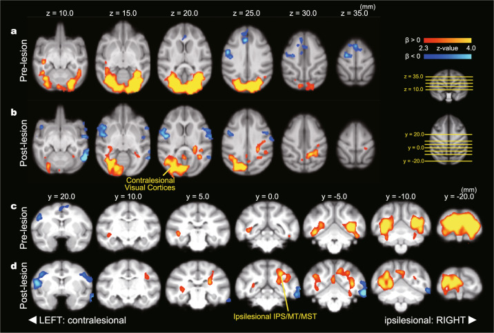
Fig. 3. Task-dependent change in rCBF in pre- and post-lesion period.
Axial (a, b) and coronal (c, d) views of PET results obtained from monkeys C and T. The left and right sides of the brain were flipped to match the side of the lesion (the lesioned side is presented on the right side in this figure). a, c Brain areas with a significant relationship to task condition in the pre-lesion period. Red-yellow and blue-light blue show a significant positive and negative relationship to task condition, respectively. Only statistically significant clusters (p < 0.05) are shown. b, d Brain areas with a significant relationship to task condition in the post-lesion period. IPS intraparietal sulcus area, MCC midcingulate cortex, MST medial superior temporal area, MT middle temporal area, SMA supplementary motor area.

