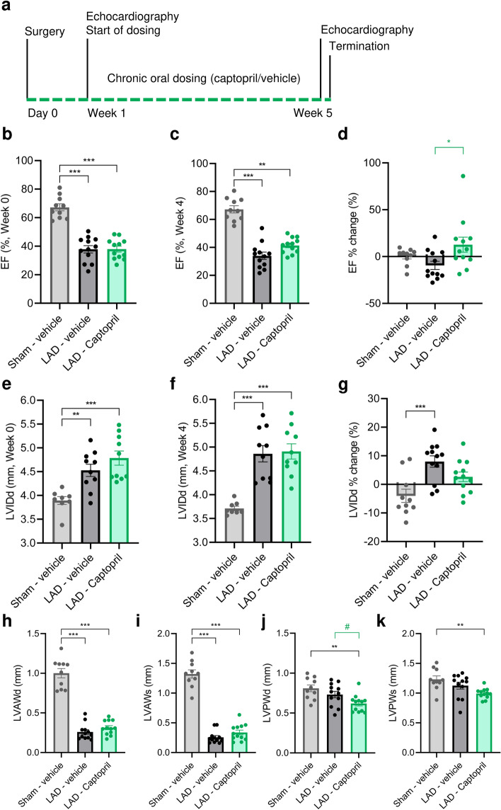
Figure 1.
Echocardiographic evaluation of cardiac function and remodelling in a mouse model of myocardial infarction. (a) Schematic study outline. (b) Ejection fraction (EF) at the time of inclusion in week 0. (c) EF after 4 weeks of dosing with either vehicle or captopril. (d) EF change over study period (week 4-week 0)/week 4*100%. (e) Left ventricle internal diameter in diastole (LVIDd) at the time of inclusion in week 0. (f) LVIDd at week 4. (g) LVIDd change over study period. (h) Left ventricle anterior wall dimension/thickness (LVAW) in diastole and (i) systole. (j) Left ventricle posterior wall dimension/thickness (LVPW) in diastole and (k) systole. Data is presented as mean ± s.e.m. n = 10–12. One-way ANOVA with Tukey’s post hoc test. Significance: *p < 0.05, **p < 0.01, ***p < 0.001. #: Significant (p < 0.05) after removal of single non-responder in LAD—Captopril. LAD: left anterior descending artery ligation.

