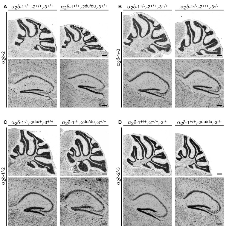
Figure 3.
General histologic examination of Nissl-stained brain sections does not reveal major morphological abnormalities. Representative micrographs of Nissl-stained sagittal cryosections obtained from adult (8–13-weeks-old; A,B) and juvenile (3–4-weeks-old; C,D) mouse brains. The cerebellum and hippocampus of ducky (A), α2δ-1/-3 (B), α2δ-1/-2 (C), α2δ-2/-3 (D) double knockout mice showed no overt anatomical defects compared to control mice. Scale bars, 400 μm (Cerebellum), and 200 μm (Hippocampus).

