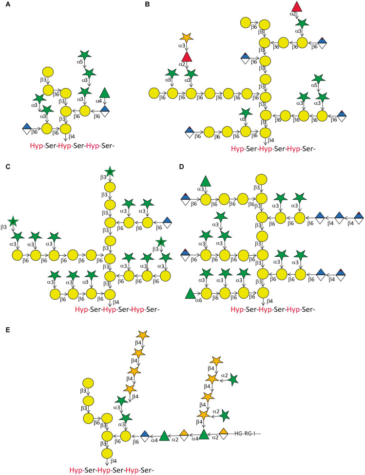
FIGURE 3.
Arabinogalactan-protein glycan variation. Five structures of type II glycans found on AGPs demonstrating common motifs and variations. (A) This relatively small glycan was produced on an artificial AGP recombinantly expressed in tobacco cell cultures by Tan et al. (2004). Note the β-(1 → 6) kink in the β-(1 → 3) galactan backbone. (B) This structure approximates the model described for AGP glycans purified from A. thaliana leaves (Tryfona et al., 2012). Note that the actual size of many of the glycans is probably much bigger than the structure displayed here. In a later study by the same group, the terminal modification of L-Fuc by D-Xyl was described (Tryfona et al., 2014). (C) Using the same tools of enzymatic degradation and mass spectrometry, this group also described the glycan-structure of wheat flour AGP (Tryfona et al., 2010). Again, we show an approximation of their model that should accommodate large variations in glycan size. A noteworthy feature of this glycan is the occurrence of terminally linked L-Arap. (D) AGP-glycans of the see grass Zostera marina are particularly rich in 4-Me-GlcAp (Pfeifer et al., 2020). (E) The partial glycan structure of the type II AG linked to an AGP named as APAP1 that is linked to both rhamnogalacturonan 1 (RG1) and arabinoxylan (AX) (Tan et al., 2013). Legend for sugar symbols is as per Figure 2.

