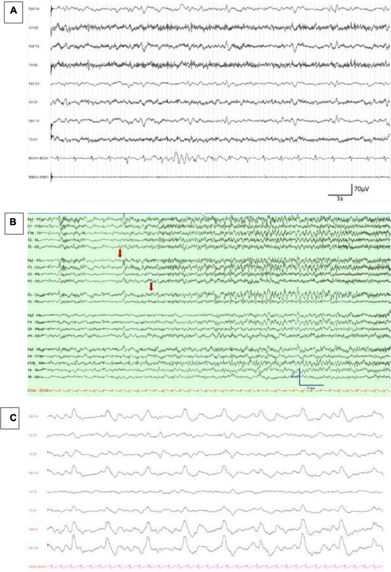
FIGURE 1.
EEG findings in COVID-19 patients. (A) Diffuse theta–delta slowing and continuous generalized periodic discharges, reproduced with authors’ agreement from Petrescu et al. (2020). (B) Emergence of low-amplitude ictal fast rhythmic activity over left frontocentral and midline regions (marked with an arrow), reproduced with authors’ agreement from Somani et al. (2020). (C) Continuous, periodic, monomorphic diphasic, delta slow waves over both frontal areas, published in Vellieux et al. (2020).

