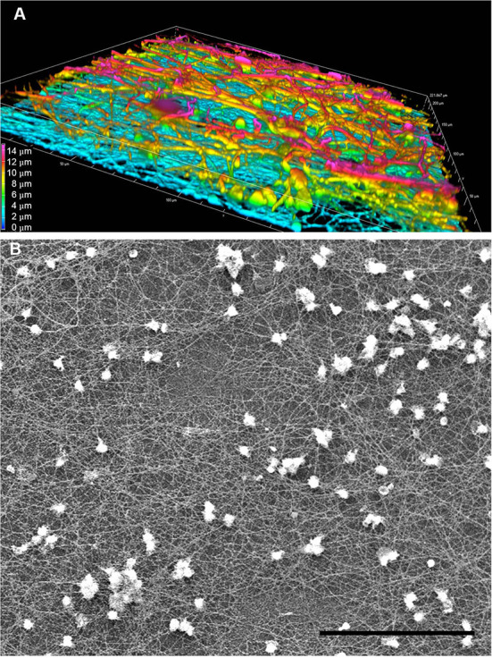
Figure 5.

Complexity and heterogeneity of LPS-induced TNTs in PHM culture. Representative images obtained by (A) 3D confocal spinning disk IF with anti-α-tubulin antibody demonstrate multilayered nature of PHM-derived TNTs and (B) SEM at low magnification (500x) demonstrates large field depth of the TNT web. (A) shows a volume view with maximum intensity projection color-coded by depth, as produced by the NIS Elements software package. Imaging microscopy performed as described in Materials and Methods (Microscopy Techniques: spinning disk confocal microscopy and high resolution SEM, respectively). Scale bar is 15 μm (A) and 100 μm (B).
