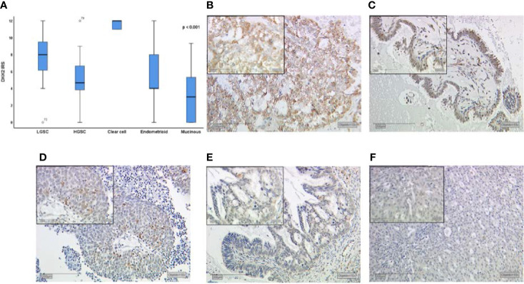
Figure 1.
Dkk2 expression patterns in different histological subtypes of EOC after immunohistochemical staining was performed as shown in a Kruskal-Wallis analysis for histological subtypes (A). Clear cell carcinomas (B) presented the strongest staining patterns. Low-grade serous carcinomas (LGSC; C) had shown moderate Dkk2 expression. For endometrioid (D), mucinous (E) and high-grade serous carcinomas (HGSC; F) the median IRS was lower. Scale bares equal 200 μm.

