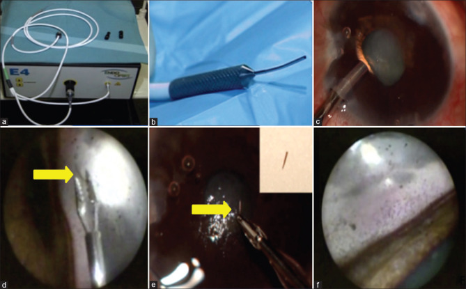
Figure 2.
(2a, 2b) Console and probe of E4 laser and Endoscopy system (2c) Insertion of endoscopic probe at diametrically opposite end of embedded bee sting (2d) Endoscopic view of the stinger (yellow arrow). A serrated 23 G forceps was used for grasping the stinger and it was subsequently removed and delivered out. The removed stinger (yellow arrow) can be seen clearly in inset (2e). (2f) show endoscopy images of anterior chamber angle clearly delineating structures of the angle

