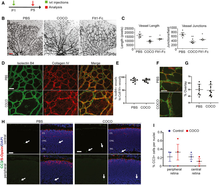
Schematic of the experimental strategy to assess early formation of the retinal vasculature (P1–P5).
Retinal flat mounts of P5 mice injected with PBS, COCO, or Flt1Fc are stained with IB4 (negative images of the fluorescent signal). Scale bar, 100 μm.
Quantification of vascular length and number of branchpoints (P‐values are vs. PBS treatment). Vessel length: **P = 0.0059 (PBS vs. COCO); *P = 0.0169 (PBS vs. Flt1Fc). Vessel junctions: **P = 0.0063 (PBS vs. COCO); *P = 0.0159 (PBS vs. Flt1Fc); (n = 4 mice/group).
COCO does not increase empty collagen IV sleeves. Scale bar, 50 μm.
Quantification of % of IsoB4 + vessels to Coll IV + vessels; (n = 9).
Visualization of pericyte coverage (NG2:green; IsoB4:red) in PBS‐ or COCO‐injected retinas. Scale bar, 50 μm.
Quantification of percentage of IsoB4 vascular staining covered by NG2 staining (% overlay) (n = 8).
COCO injections (P1) do not result in apoptosis in P5 retinas. Scale bar, 40 μm.
Quantification of apoptotic cells in control and COCO‐injected mice (n = 3). Results are presented as mean ± SEM and statistical significance was analyzed by Mann–Whitney test. *P < 0.05, **P < 0.01.

