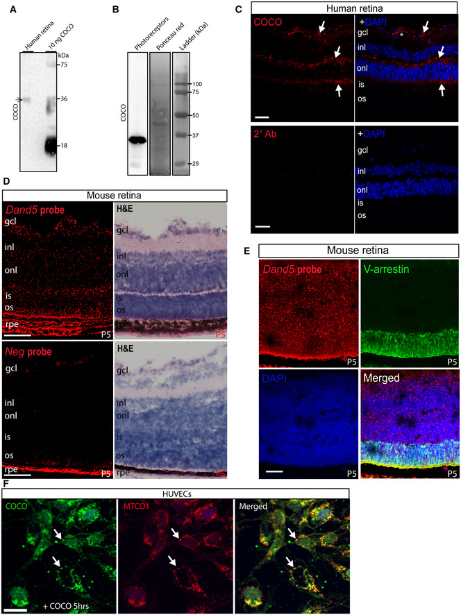
Figure 7. COCO is expressed in human and mouse retina and localizes to mitochondria in COCO‐exposed HUVECs.

- Western blot of whole adult human retina extracts incubated with an anti‐human COCO antibody and revealing a unique band at ~ 36 kDa (arrow). Recombinant human COCO was used as positive control.
- Western blot of photoreceptors produced from human embryonic stem cells incubated with an anti‐mouse COCO antibody and revealing a unique band at ~ 36–38 kDa (arrow). Western blots are representative of 3 independent experiments.
- Immunofluoresence analysis of adult human retina sections with an anti‐mouse COCO antibody. Specific immunoreactivity was observed in multiple areas (arrows) when compared with sections only exposed to the secondary antibody. Scale bar, 40 μm.
- RNAscope in situ hybridization (Dand5, top; Negative probe (dapB); down) and hematoxylin staining of P5 mouse retinas. Scale bar, 40 μm.
- Dual RNAscope in situ hybridization and visual‐arrestin immunohistochemistry of P5 mouse retinas. Scale bar, 40 μm. Images are representative of 4 animals.
- Immunofluoresence analysis of HUVECs exposed to COCO for 5 h prior to fixation. Exogenously added COCO was detected using an anti‐human COCO antibody, showing co‐localization with human mitochondria (MTCO1 antibody). Scale bar 25 μm.
